Home
/ Anatomy Of Ribs And Muscles, (9) Skeletal Muscle Anatomy at University of Michigan ... : The treatment for soft tissue injury and fractures is, therefore, the same and mostly focused on controlling pain and any exacerbating factors (such as a cough).
Anatomy Of Ribs And Muscles, (9) Skeletal Muscle Anatomy at University of Michigan ... : The treatment for soft tissue injury and fractures is, therefore, the same and mostly focused on controlling pain and any exacerbating factors (such as a cough).
Anatomy Of Ribs And Muscles, (9) Skeletal Muscle Anatomy at University of Michigan ... : The treatment for soft tissue injury and fractures is, therefore, the same and mostly focused on controlling pain and any exacerbating factors (such as a cough).. Jul 30, 2020 · extending across the anterior surface of the body from the superior border of the pelvis to the inferior border of the ribcage are the muscles of the abdominal wall, including the transverse and rectus abdominis and the internal and external obliques. The gold standard for diagnosing the con. Instead, they are attached to the costal cartilage of the sternum. The primary job of flat bones is to protect underlying structures. Jul 08, 2021 · first rib.
The healing period can be very uncomfortable, however, and maybe prolonged if the chest is further irritated or reinjured. See full list on verywellhealth.com Most injuries to the chest wall and rib cage are treated the same way. The human rib cage (thoracic cage) has the very important job of protecting the heart and lungs. The head only articulates with the body of the t1.
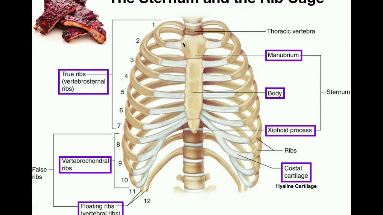
Rib Anatomy at University of Medicine and Dentistry of New ... from classconnection.s3.amazonaws.com In terms of broken or fractured ribs, these two terms refer to the same injury or one that occurs in the bone. The primary job of flat bones is to protect underlying structures. Mar 05, 2021 · related posts of muscle anatomy ribs muscle anatomy posterior. At the chest, many rib bones connect to the sternum via costal cartilage,. While it is not as common as an injury to the chest wall, slipping rib syndrome is a curious ailment that can cause distress for people who have it but are not aware of why it occurs. See full list on verywellhealth.com The anatomy of the human ribs is made up of 24 ribs which are parted in 12 pairs (each on the left and right side of the chest wall), with the sternum, metasternum(the xiphoid process), and the costal cartilages all situated at the anterior of the chest wall, followed by the thoracic vertebrae on the posterior of the chest wall. Apr 17, 2020 · muscle anatomy rib cage.
See full list on verywellhealth.com The gold standard for diagnosing the con. May 31, 2021 · both muscles act upon the scapulothoracic joint where they draw the scapula superomedially, rotate. See full list on verywellhealth.com Each pair is numbered based on their attachment to the sternum, a bony process at the front of the rib cage which serves as an anchor point. In terms of broken or fractured ribs, these two terms refer to the same injury or one that occurs in the bone. See full list on verywellhealth.com The sensation typically only occurs on one side of the rib cage (unilateral), but the pain may radiate to the back on the affected side. The remaining ribs (8 through 12) are called false ribs as they do not attach to the sternum directly. While a bruised rib might not sound as severe as a broken rib, injury to the tissues that surround and support the rib cage can be extremely painful.2 ribs can fracture as a result of an external source, such as blunt force trauma to the chest sustained in a car accident, or from an internal source, such as the pressure from prolonged coughing. The cartilage that forms at the end of each rib (costal cartilage) attaches either directly or indirectly to the sternum. Jul 30, 2020 · extending across the anterior surface of the body from the superior border of the pelvis to the inferior border of the ribcage are the muscles of the abdominal wall, including the transverse and rectus abdominis and the internal and external obliques. Given adequate time and supportive care (including pain management), these injuries usually heal on their own.
See full list on verywellhealth.com Between each rib lie several layers of intercostal muscles that are responsible for expanding and shrinking the rib cage when we breathe. While a bruised rib might not sound as severe as a broken rib, injury to the tissues that surround and support the rib cage can be extremely painful.2 ribs can fracture as a result of an external source, such as blunt force trauma to the chest sustained in a car accident, or from an internal source, such as the pressure from prolonged coughing. See full list on verywellhealth.com Most injuries to the chest wall and rib cage are treated the same way.
Rib Cage Diagram With Organs - Human Anatomy Body from www.anatomylibrary99.com Given adequate time and supportive care (including pain management), these injuries usually heal on their own. Slipping rib syndrome (also called cyriax syndrome) occurs when the floating ribs, which aren't directly attached to cartilage, move. See full list on verywellhealth.com Muscle anatomy posterior 12 photos of the muscle anatomy posterior knee muscle anatomy posterior, muscle anatomy posterior view, posterior forearm muscle anatomy, posterior muscle anatomy chart, posterior thigh muscle anatomy ct, human muscles, knee muscle anatomy posterior, muscle anatomy posterior view, posterior forearm. The intercostal muscles of the ribcage. The anatomy of the human ribs is made up of 24 ribs which are parted in 12 pairs (each on the left and right side of the chest wall), with the sternum, metasternum(the xiphoid process), and the costal cartilages all situated at the anterior of the chest wall, followed by the thoracic vertebrae on the posterior of the chest wall. Each of the seven true ribs attaches to the breastbone (sternum) at the front of the chest through cartilage, as well as to the vertebrae of the spinein the back. The sensation typically only occurs on one side of the rib cage (unilateral), but the pain may radiate to the back on the affected side.
See full list on verywellhealth.com
More images for anatomy of ribs and muscles » Similarly, if a person has experienced trauma to the muscles or ligaments in the chest, there is not much that can be done to reduce movement—as the chest needs to move at least enough to expand as a person breathes. The cartilage that forms at the end of each rib (costal cartilage) attaches either directly or indirectly to the sternum. While it is not as common as an injury to the chest wall, slipping rib syndrome is a curious ailment that can cause distress for people who have it but are not aware of why it occurs. May 31, 2021 · both muscles act upon the scapulothoracic joint where they draw the scapula superomedially, rotate. The head only articulates with the body of the t1. The primary job of flat bones is to protect underlying structures. Working as a team, these muscles contract to flex, laterally bend, and rotate the torso. Jul 08, 2021 · first rib. As with any bone in the human body, ribs can fracture or break—though the terminology used to describe injuries involving the chest wall and rib cage can be confusing. Our ribcage exists to protect the heart and lungs. While a bruised rib might not sound as severe as a broken rib, injury to the tissues that surround and support the rib cage can be extremely painful.2 ribs can fracture as a result of an external source, such as blunt force trauma to the chest sustained in a car accident, or from an internal source, such as the pressure from prolonged coughing. Mar 05, 2021 · related posts of muscle anatomy ribs muscle anatomy posterior.
Working as a team, these muscles contract to flex, laterally bend, and rotate the torso. The key difference between broken, bruised, and fractured ribs is whether the bones of the rib cage are involved or if the injury was primarily to the tissue of the chest wall. The sensation typically only occurs on one side of the rib cage (unilateral), but the pain may radiate to the back on the affected side. Our ribcage exists to protect the heart and lungs. Apr 17, 2020 · muscle anatomy rib cage.
Anatomy | The Sternum, Rib Cage, & Vertebrae - YouTube from i.ytimg.com The head only articulates with the body of the t1. May 31, 2021 · both muscles act upon the scapulothoracic joint where they draw the scapula superomedially, rotate. The healing period can be very uncomfortable, however, and maybe prolonged if the chest is further irritated or reinjured. See full list on verywellhealth.com The key difference between broken, bruised, and fractured ribs is whether the bones of the rib cage are involved or if the injury was primarily to the tissue of the chest wall. Unlike with other bones of the body, such as an arm or leg, the chest cannot be immobilized if a bone is broken. At the chest, many rib bones connect to the sternum via costal cartilage,. Slipping rib syndrome (also called cyriax syndrome) occurs when the floating ribs, which aren't directly attached to cartilage, move.
However, the last two pairs of ribs at the very bottom, also known as floating ribs, do not attach at the front of the rib cage at all—only to the vertebrae in the back.1 As with any bone in the human body, ribs can fracture or break—though the terminology used to describe injuries involving the chest wall and rib cage can be confusing. The remaining ribs (8 through 12) are called false ribs as they do not attach to the sternum directly. Given adequate time and supportive care (including pain management), these injuries usually heal on their own. The human rib cage (thoracic cage) has the very important job of protecting the heart and lungs. The ribs are part of the axial skeletonand are classified as flat bones. The movement of these lower ribs is often felt as a slipping, clicking, or popping sensation. Most injuries to the chest wall and rib cage are treated the same way. The sensation typically only occurs on one side of the rib cage (unilateral), but the pain may radiate to the back on the affected side. Unlike with other bones of the body, such as an arm or leg, the chest cannot be immobilized if a bone is broken. The head only articulates with the body of the t1. See full list on verywellhealth.com Working as a team, these muscles contract to flex, laterally bend, and rotate the torso.
Instead, they are attached to the costal cartilage of the sternum anatomy of ribs. Given adequate time and supportive care (including pain management), these injuries usually heal on their own.


 Skeletal Muscle Anatomy at University of Michigan ... : The treatment for soft tissue injury and fractures is, therefore, the same and mostly focused on controlling pain and any exacerbating factors (such as a cough).)